

[Close]
- This page was created by volunteers like you!
- Help us make it even better. To learn more about contributing to MEpedia, click here.
- Join the movement
- Visit #MEAction to find support or take action. Donate today to help us improve and expand this project.
- Congratulations!
- MEpedia has got over 30 million views as of August 2022!
File:Microglia.JPG
From MEpedia, a crowd-sourced encyclopedia of ME and CFS science and history
No higher resolution available.
Microglia.JPG (545 × 412 pixels, file size: 58 KB, MIME type: image/jpeg)
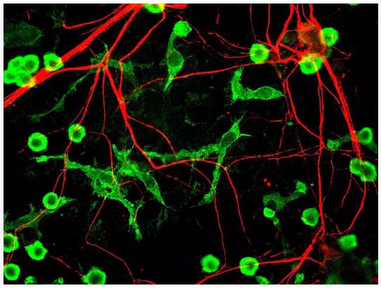
Title: Microglia and Neurons
Author: Gerry Shaw
Source: Microglia and neurons - Wikimedia
License: Attribution-ShareAlike 3.0 Unported CC BY SA 3.0
Description: Mixed rat brain cultures stained for coronin 1a, found in microglia here in green, and alpha-internexin, in red, found in neuronal processes. Antibodies and image generated by EnCor Biotechnology Inc.

|
This file is licensed under the Creative Commons Attribution-Share Alike 3.0 Unported license. | |
|
File history
Click on a date/time to view the file as it appeared at that time.
| Date/Time | Thumbnail | Dimensions | User | Comment | |
|---|---|---|---|---|---|
| current | 02:40, July 19, 2018 | | 545 × 412 (58 KB) | MEcfsFMS (talk | contribs) | Microglia Alzheimers progression (top): By National Institute on Aging [Public domain], Source: Wikimedia Commons |
You cannot overwrite this file.
File usage
The following 3 pages use this file: